
Figure 1.
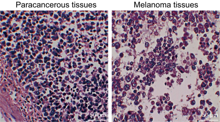
The mouse model was successfully established as evidenced by histopathological assessment in the paracancerous tissues and the melanoma tissues by haematoxylin and eosin staining (×200). HE, haematoxylin‐eosin
Official websites use .gov
A
.gov website belongs to an official
government organization in the United States.
Secure .gov websites use HTTPS
A lock (
) or https:// means you've safely
connected to the .gov website. Share sensitive
information only on official, secure websites.
The mouse model was successfully established as evidenced by histopathological assessment in the paracancerous tissues and the melanoma tissues by haematoxylin and eosin staining (×200). HE, haematoxylin‐eosin