
Figure 1.
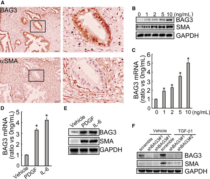
BAG3 is highly expressed in activated pancreatic stellate cells. (A) Representative immunohistochemical staining of BAG3 (upper left) and α‐SMA (upper right) in human PDAC tumour cells and stroma. The lower images show the selected boxed area of upper left. (B) HPanSteC cells were treated with TGF‐β1 (1, 2, 5 and 10 ng/mL) for 48 hours and the protein levels of BAG3 and α‐SMA were analysed by Western blot. (C) HPanSteC cells were treated with the indicated concentrations of TGF‐β1 for 24 h, the BAG3 mRNA level was detected by RT‐qPCR. (D and E) HPanSteC cells were stimulated with PDGF and IL6, mRNA level and protein level of BAG3 were analysed by RT‐qPCR and Western blotting respectively. (F) HPanSteC cells were infected with lentivirus containing shRNAs against BAG3 (shBAG3) for 48 h, then treated with 10 ng/mL TGF‐β1 for additional 24 h. Western blotting was performed to detect the protein levels of BAG3 and α‐SMA. *P < 0.01. Error bars indicate means ± SD