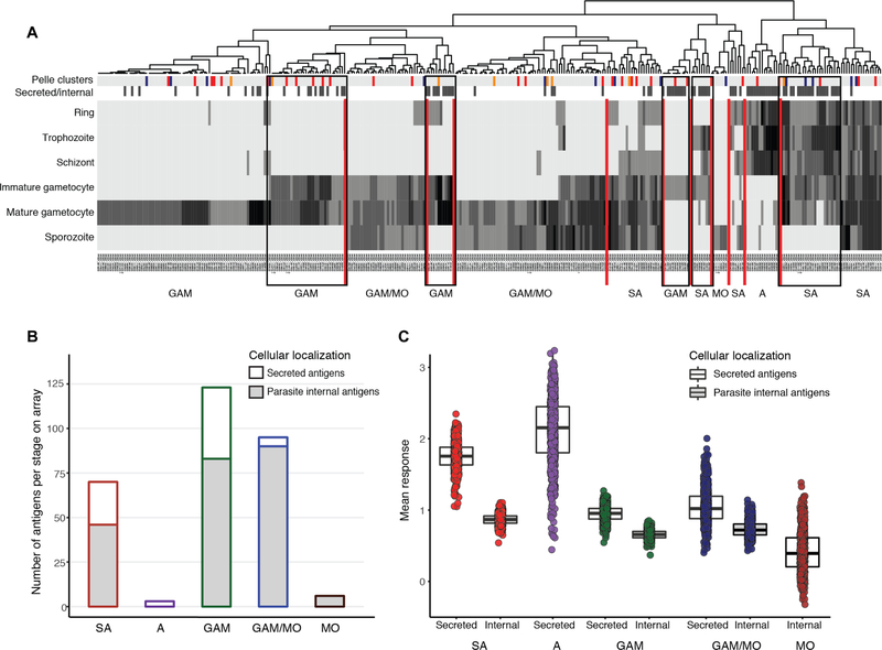
Fig. 1. Human plasma samples recognize secreted asexual (aiRBC) and gametocyte (giRBC) surface antigens.
(A) Heatmap of 344 P. falciparum antigens from 3D7 genome (PlasmoDB release 31) clustering proteins on the array by timing of protein expression (log read counts of number of peptides sequences). Additional annotations are indicated by color bars at the top of the heatmap: The first row indicates cluster stage annotation from (52) (orange, gametocyte rings; red, immature gametocytes; blue, mature gametocytes; gray, others), and the second row indicates cellular localization (black, secreted; white, internal/unknown). Vertical red lines separate stage-specific clusters. Black boxes highlight five clusters of shared or gametocyte-specific secreted antigens. (B) Distribution of 528 P. falciparum protein fragments on the peptide array [developed in (22)] by stage and location. The proteins were selected on the basis of expression during gametocyte stages and predicted export (details in table S2). (C) Mean responses across three malaria-exposed populations are quantified by peptide array (after normalization to controls and quantile normalization), stage of protein expression, and whether they are secreted or not (see table S1). GAM, gametocyte; GAM/MO, gametocyte/mosquito stages; MO, mosquito stages; A, asexual stages; SA, shared antigens.

