
Figure 1.
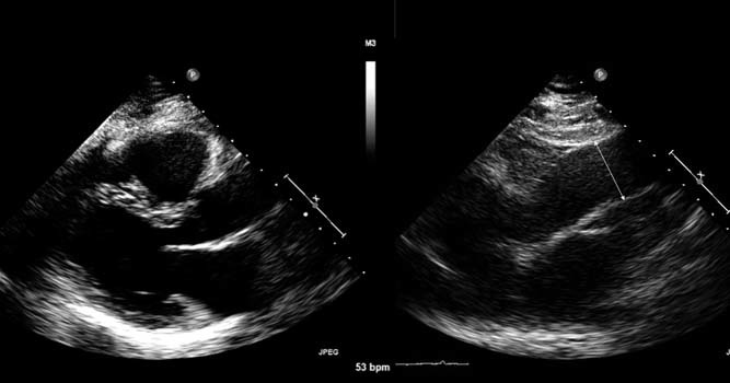
Left panel : Standard transthoracic parasternal long‐axis image of the aortic root. Right panel : Transthoracic parasternal long‐axis image with movement up an interspace for visualization of the ascending aorta and measurement of the inner wall to inner wall of the tubular ascending aorta