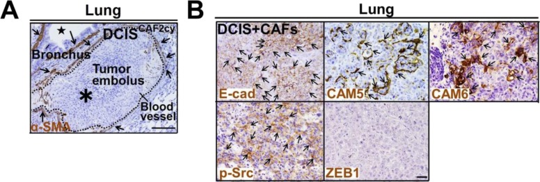
Figure S7. Related to Fig 8. CAF-induced CTC clusters, tumor emboli, and metastatic colonization.
(A) Immunohistochemistry of lung sections prepared from mice at 60 d after subcutaneous injection of DCISCAF2cy using anti–α-SMA antibody. Tumor embolus (asterisk in a broken circle) is indicated. α-SMA–positive cells (arrows) are detected in smooth muscle cell layers in the blood vessel and bronchus (star). (B) Immunostaining of lung sections prepared from mice at 60 d after subcutaneous injection of DCIS cells admixed with CAFs, using the indicated antibodies. Note that DCIS cells show highly positive staining for E-cad, CAM5, CAM6, and p-Src (arrows), in contrast to negative staining for ZEB1. Data information: Scale bars, 100 μm (A) and 30 μm (B).

