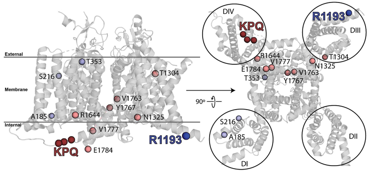
Figure 5:
Cartoon representation of a model of NaV1.5 highlighting the membrane spanning segments as well as R1193 and residues 1505-1507 (KPQ, removed in ΔKPQ). Rightmost image is from the perspective of inside the cell looking out through the channel and membrane. R1193 is in the amphipathic helix preceding the transmembrane domain of domain III while ΔKPQ is in the domain III-IV linker, the segment of SCN5A known to influence inactivation.33 The model is based on electric eel NaV1.4 (67% sequence identity to human NaV1.5) structure (PDBID: 5XSY).22 The four helix voltage-sensing modules of each domain (I-IV) are noted by black circles. Residues where variants with at least five carriers and late current greater than 200% that of WT are known are shown as spheres. Light blue spheres are variants where the predicted penetrance is < 20%; light red spheres are variants where predicted penetrance is > 20%. The high-penetrance variants are spatially proximal to the inactivation machinery, in contrast with low-penetrance variants.

