
Fig. 3.
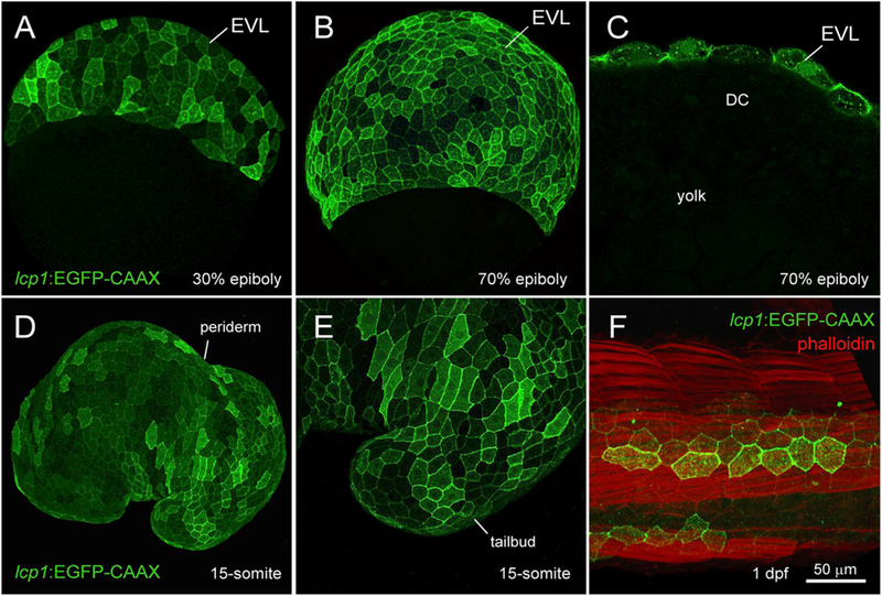
Transgenic expression in lcp1:EGFP-CAAX embryos.
A. 30% epiboly, transgenic embryo in equatorial view. The EVL shows weak, variegated EGFP expression.
B. 70% epiboly, transgenic embryo in equatorial view. Signal intensifies as the EVL spreads over the yolk.
C. 70% epiboly, cutaway view of a single focal plane. Only EVL cells are EGFP +, in contrast to the deep cells (DC) and yolk syncytial layer (YSL).
D. Live embryo at the 15-somite stage. lcp1:EGFP-CAAX labels the periderm, the outermost epithelial layer.
E. Magnification of the tailbud in D, showing variegated EGFP in adjacent peridermal cells.
F. Fixed embryo at 1 dpf, with phal-loidin-568 counterstain. A patch of label-retaining peridermal cells (green) lies over the developing trunk musculature (red).