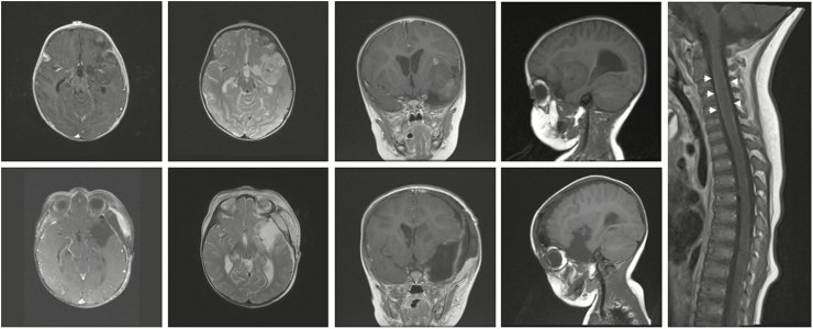
Fig. 1.
Imaging Findings in a Patient With an Atypical Teratoid/Rhabdoid Tumor. Top row: Preoperative MRI demonstrated a heterogeneous, patchily enhancing left frontotemporal mass encasing branches of the middle cerebral artery with minimal surrounding cerebral edema. Right panel: Whole-spine MRI showed diffuse leptomeningeal enhancement of the spinal cord (arrowheads). Bottom row: Postoperative MRI demonstrated interval gross total resection of the mass of the tumor with improved mass effect.

