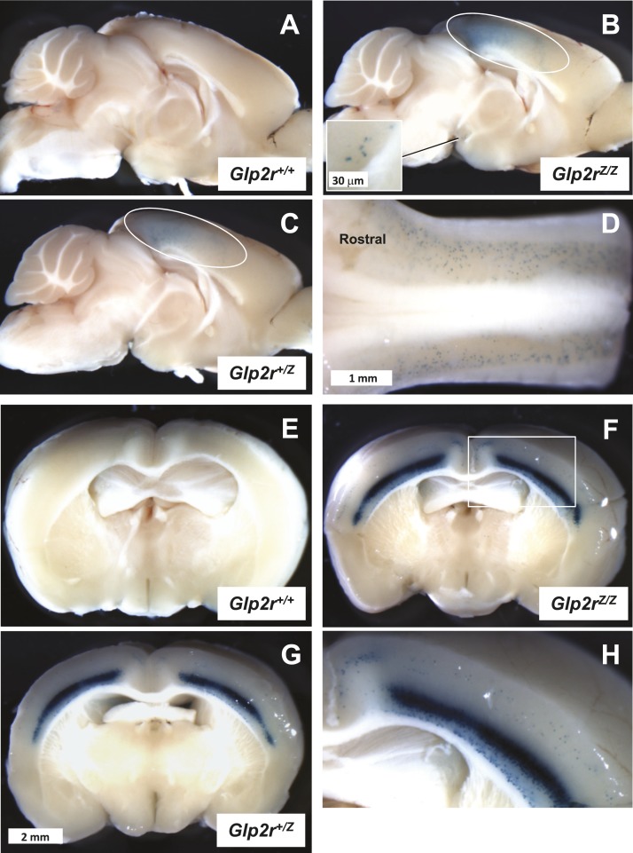
Figure 10.
Whole-mount (A–C) midsagittal and (E–H) coronal sections from (A and E) Glp2r+/+, (C and G) Glp2r+/Z, and (B, F, and H) Glp2rZ/Z mouse brain following histochemical detection of β-galactosidase activity. (A–C) All images are shown at the same magnification, as they are in (E–G). (D) Dorsal view of the Glp2rZ/Z mouse brainstem demonstrating the presence of β-galactosidase-positive cells in the nucleus of the solitary tract. The cerebellum has been removed to visualize the brainstem. (B and C) The area enclosed by the ovals outlines β-galactosidase positivity in the region of the visual and somatosensory cortex. (B) The inset illustrates a few scattered β-galactosidase–positive cell bodies noticeable in the hypothalamus. (H) A close view of the Glp2rZ/Z mouse brain coronal section of (F), inset, featuring strong β-galactosidase activity in the inner layers of the somatosensory cortex. Images are representative of independent whole-mount preparations from two Glp2r+/+, three Glp2r+/Z, and two Glp2rZ/Z mice.

