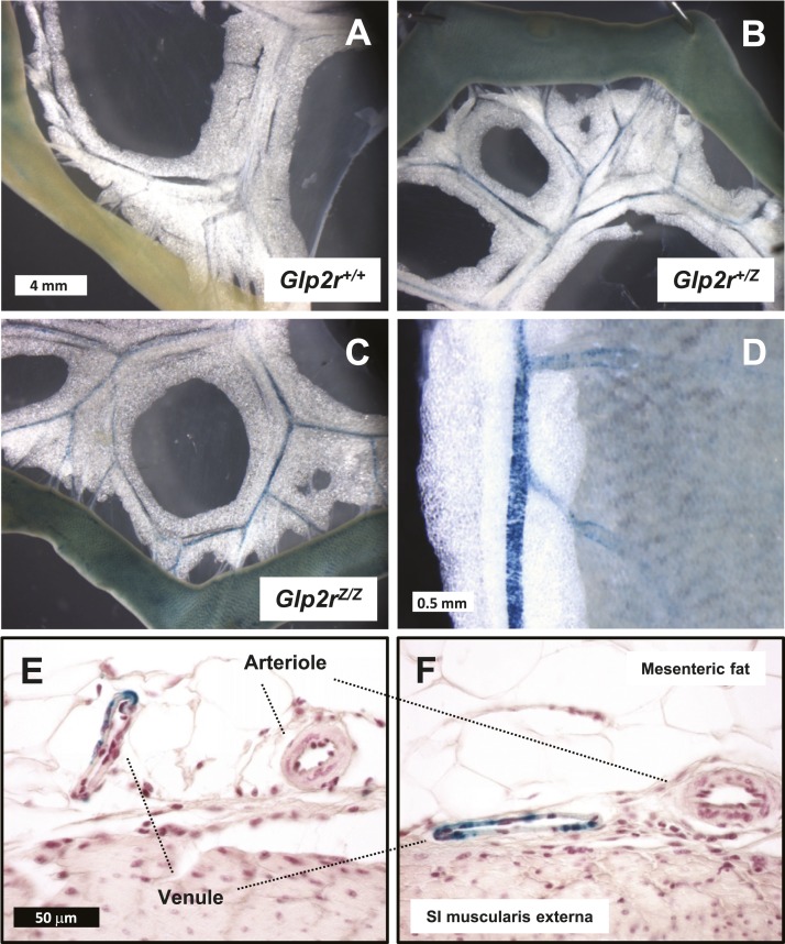
Figure 9.
(A–D) Whole mounts and (E and F) histological sections from (A) Glp2r+/+, (B) Glp2r+/Z, and (C–F) Glp2rZ/Z mouse mesenteric vascular bed following histochemical detection of β-galactosidase activity. (D) Higher magnification of a whole mount from a Glp2rZ/Z mouse mesenteric blood vessel embedded in mesenteric fat. (A–C) All images are shown at the same magnification. (E and F) Histological sections are at the same scale. Images are representative of independent whole-mount preparations from two Glp2r+/+, three Glp2r+/Z, and two Glp2rZ/Z mice and multiple histological tissue sections from two Glp2rZ/Z mice. SI, small intestine.

