Figure 6.
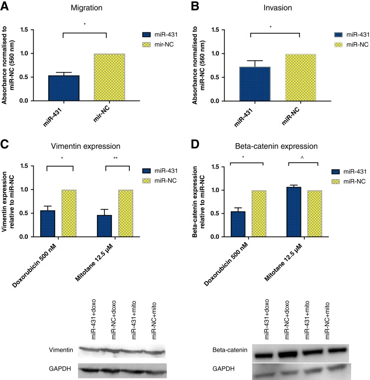
The effects of miR‐431 on epithelial‐mesenchymal transition processes. (A): Following miR‐431/NC restoration, cells were treated with 500 nM doxorubicin for 48 hours and assessed for migration via transwell assay. There was a 46% decrease on migration in miR‐431‐restored cells. Error bars represent SEM. *, p = .02. (B): Following miR‐431/NC restoration, cells were treated with 500 nM doxorubicin for 48 hours and assessed for invasion via transwell assay. There was a 27% decrease on invasion in miR‐431‐restored cells. Error bars represent SEM. *, p = .01. (C): Following miR‐431/NC restoration, cells were treated with 500 nM doxorubicin or 12.5 μM mitotane for 48 hours. Vimentin was twofold decreased in miR‐431‐restored cells treated with doxorubicin or mitotane. Error bars represent SEM, n = 3. *, p = .007. **, p = .04. Representative images of one Western blot experiment are shown. (D): Following miR‐431/NC restoration, cells were treated with 500 nM doxorubicin or 12.5 μM mitotane for 48 hours. In miR‐431 + doxorubicin cells, there was a twofold decrease in beta‐catenin expression. This was not detected for miR‐431 cells treated with mitotane. Error bars represent SEM, n = 3. *, p = .008. ^, p = .09. Representative images of one Western blot experiment are shown.
Abbreviations: doxo, doxorubicin; GAPDH, glyceraldehyde 3‐phosphate dehydrogenase; miR‐NC, negative control; mito, mitotane.

