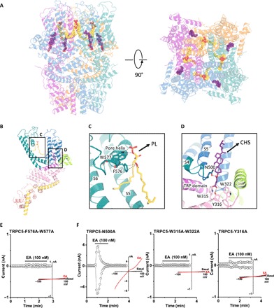
Fig. 6. Lipid coordination in TRPC5.

Lipid-channel interactions. (A) Side and top views of ribbon diagrams of the TRPC5 tetramer: four CHS molecules and four PLs (potentially ceramide-1-phosphate or phosphatidic acid) are shown as spheres with purple or yellow carbons, respectively. (B) Side views of each CHS and PL molecules per protomer. (C and D) Ribbon diagram of the TRPC5 lipid binding regions. (C) PL interacts with the pore helix through Trp577 and Phe576. (D) CHS, shown in purple, interacts with the S4/S5 linker at Asn500 and the N-terminal domains at Trp315, Tyr316, and Trp322. (E and F) Patch clamp recordings of lipid binding site mutants in response to EA.
