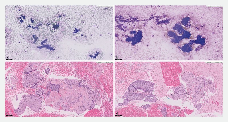
Fig. 4.

ROSE and cell-block assessment of samples obtained from a lymph node in metastatic squamous cell carcinoma. a Diff-Quick staining of cytology specimen obtained using Franseen EUS-FNB. b Diff-Quick staining of similar cellular yield obtained using EUS FNA. c H&E staining of histology obtained using Franseen EUS-FNB shows malignant cells surrounded by dense desmoplastic stroma. d H&E staining of histology obtained using EUS-FNA shows malignant cells with scanty desmoplastic stroma.
