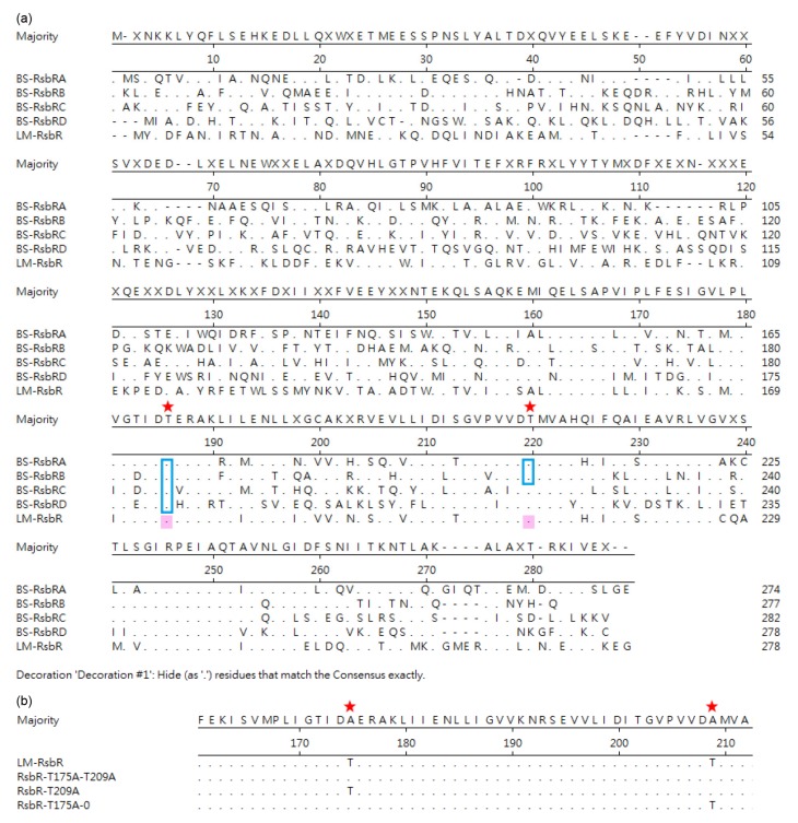
Fig. 1.
Sequence alignment of RsbR homologs of Bacillus subtilis (BS) and Listeria monocytogenes (LM) (a) and alanine substitutions in the recombinant complementary plasmids (b)
Blue squares indicate the phosphorylation sites in PhosphositePlus (https://www.phosphosite.org); pink shading indicates the phosphorylation sites predicted by DISPHOS (http://www.dabi.temple.edu/disphos); red stars indicate the conservative phosphorylation sites in both BS and LM (Note: for interpretation of the references to color in this figure legend, the reader is referred to the web version of this article)

