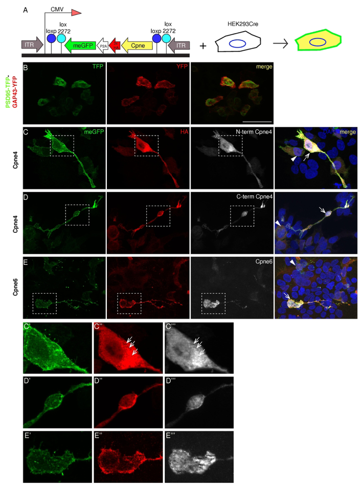
Figure 4. Sub-cellular localization of Copines 4 and 6 in HEK293 cells.
A, Expression construct and strategy. The expressed cDNA consists of the Cpne gene tagged with 3x HA, self-cleaving P2A signal and eGFP coupled to a GAP43 membrane localization signal (meGFP). The entire cassette is placed in reverse orientation between the tandem inverted lox sites- loxp and lox 2272 downstream of a CMV promoter. Transfection into HEK293Cre cells leads to an irreversible inversion of the expression cassette and two fusion proteins are synthesized- meGFP which localizes on the cell membrane and Cpne-3xHA peptide. B, Control transfection with a bicistronic construct containing PSD95-TFP and GAP43-YFP shows the lack of colocalization of the two peptides. C-E, Constructs containing Cpne4 or 6 genes were transfected into the HEK293-Cre cells. Localization of the two fusion proteins is revealed by immunostaining for GFP (green, first column), HA (red, second column) and Cpne (white, third column) antibodies indicate their localization in the HEK293Cre cells. C’-E’’’, The dashed boxes in C, D and E are zoomed in to show the differences in sub-cellular distribution between Cpne4 and 6, respectively. The meGFP is localized to the cell membrane in all the transfected cells. HA immunostaining indicates the presence of Copines in the cytosol as well as the nuclei of the cells for Cpne4 and is mostly punctate and cytosolic for Cpne6. n= 4 each for Cpne4 and 6, n=2 for control. Scale bar in B: 50μm.

