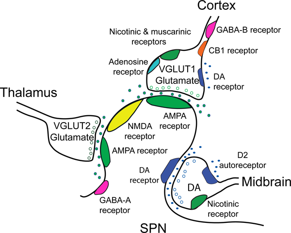
Figure 2.

The corticostriatal network and the effect of dopamine on corticostriatal terminals. The simplified cartoon shows the tripartite configuration of a glutamatergic corticostriatal input, a thalamostriatal input, and a dopaminergic (DA) midbrain input as they synapse on an SPN. Glutamatergic presynaptic elements generally display an asymmetric synaptic density with docked synaptic vesicles. The corticostriatal density contains VGLUT1 and docks near the spine head, while the thalamostriatal element contains VGLUT2 and docks on the spine shaft. A dopamine terminal is generally associated with the spine neck and shaft and displays a relatively small symmetric synaptic density (Totterdell and Smith 1989). Virtually all striatal synapses are within 2 μm of an apparent dopamine axonal varicosity, and thus are thought to receive dopamine input. SPNs express AMPA, NMDA and GABA-A receptors, in addition to D1 or D2 receptors (Wang and others 2012). Presynaptic afferents from the cortex express acetylcholine nicotinic and muscarinic receptors, adenosine and cannabinoid CB1 receptors and GABA-B receptors (Wang and others 2013a; Wang and others 2012; Wang and others 2013b). In the dorsal and ventral striatum, dopamine D2 receptors are found on presynaptic afferents to D2-SPNs (Bamford and others 2004b; Wang and others 2012). In the ventral striatum, D1-SPNs are expressed on presynaptic afferents to both D1- and D2-SPNs (Wang and others 2012). Presynaptic receptors on excitatory glutamatergic afferents from the thalamus have not been well characterized.
