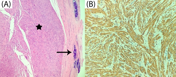
Fig. 3.
(A) Microscopic histological image showing interlacing bundles of smooth muscle fibres without atypia or mitosis (star) with urothelial ducts of posterior wall of the distal urethra (arrow). Hematoxylin-eosin staining x100. (B) Positive staining for desmin, specific marker of smooth muscle, x400.

