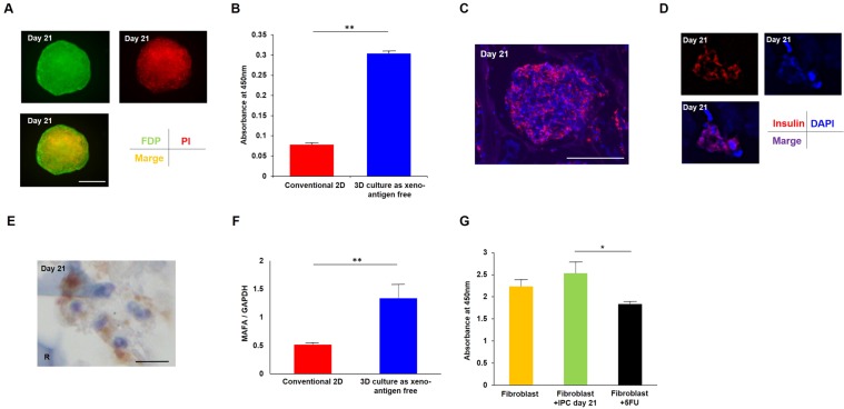
Figure 2.
Cell quality of IPCs generated by the 3D cultured protocol was better than IPCs from conventional monolayer culture. (A) The cell viability of 3D cultured IPCs at day 21 was determined by FDP/PI staining. Green: FDP staining, red: PI staining, orange: merged image. Scale bar, 100 μm. (B) Absorbance at 450 nm showed significantly better cell viability of 3D cultured IPCs compared with IPCs from conventional 2D culture method. **P < 0.01, un-paired t-test. (C) Immunofluorescence of generated IPCs at day 21. Red: insulin, blue: DAPI. Scale bar, 100 μm. (D) The larger image shows the insulin-positive area was the cytoplasm. Red: insulin, blue: DAPI. (E) Light microscopy analysis also showed the cytoplasm of generated IPCs was positive for insulin staining. Scale bar, 10 μm. R: RCP petaloid μ-piece, stained blue. (F) MAFA mRNA expression was significantly higher in 3D cultured IPCs compared with IPCs from conventional 2D culture method by unpaired t-test. **P < 0.01. Error bar indicates standard deviation. (G) Generated IPCs did not show significant adverse effects on human dermal fibroblasts in cell toxicity, although 100 μM 5-FU addition showed significantly reduced viability. *P < 0.05, one-way ANOVA.

