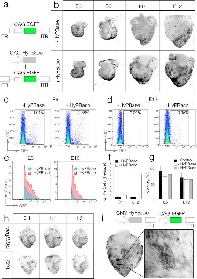
Figure 2.
Stable expression of exogenous expression cassettes. (A) Diagram of expression cassettes tested. (B) Time series of GFP expression (black) in hearts transfected with or without HyPBase enzyme. Images were grayscaled and inverted. (C) Flow cytometry data from dissociated E6 hearts (88 hrs. post transfection). Plots are presented as Forward Scatter vs GFP intensity. (D) As in (C), for hearts dissociated at E12 (240 hrs. post transfection). (E) Quantification of cell counts vs GFP intensity from (C) and (D). Note: both the number of GFP positive cells and the intensity of GFP drops in E12 hearts that were not transfected with HyPBase. (F) Relative number of GFP positive cells with and without HyPBase cotransfection (n = 3 hearts per group). (G) Percent viability among windowed control embryos, embryos transfected without HyPBase and embryos transfected with HyPBase. (H) Comparison of E9 hearts transfected with different ratios of transposase to transposable element using both the PiggyBac and Tol2 transposase systems. (I) Heart transfected with a single plasmid containing both a HyPBase and integrating EGFP expression cassette.

