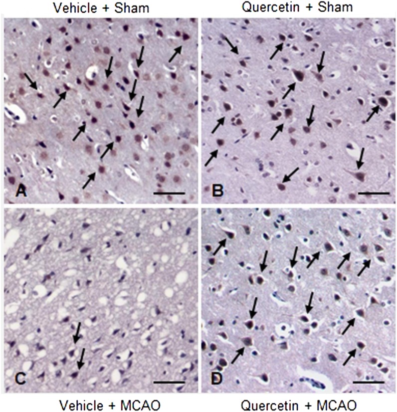
Fig. 5.
Representative photos of immunohistochemical staining of protein phosphatase 2A (PP2A) subunit B in the cerebral cortices of vehicle + sham (A), quercetin + sham (B), vehicle + middle cerebral artery occlusion (MCAO) (C) and quercetin + MCAO (D) animals. Arrows indicate positive cells of PP2A subunit B. Scale bar=100 µm.

