Figure 5.
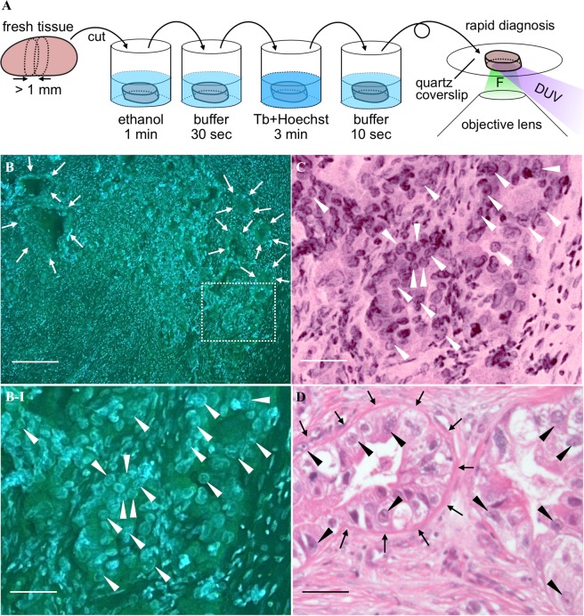
Preclinical demonstration of DUV-excitation fluorescence microscopy with Tb3+ and Hoechst for intraoperative rapid diagnosis of lymph node metastasis. (A) A schematic view representing the protocol of intraoperative rapid diagnosis of a surgical specimen, drawn by Y.K. Because the lymph node is less contaminated by blood, the tissue is not rinsed before the ethanol treatment, unlike the staining protocol presented in Methods, so that the treatment time is shorten. F: fluorescence. (B) A cancer-metastasized lymph node stained with the D2O solution containing Tb3+ (50 mM) and Hoechst (20 µg/ml). The excitation wavelength, treatment period, and solution pH were adjusted to 285 nm, 3 min, and 7, respectively. The region indicated by the dotted square is magnified in (B-I). (C) The virtual H&E image corresponding to (B-I). (D) H&E image of the correspondent lymph node sample. Scale bars correspond to 200 and 50 µm for the large field-of-view (B) and magnified (B-I,C,D) images, respectively. For the images shown in (B,B-I) unsharp masking was applied. The correspondent original images are shown in Fig. S2. Arrows indicate boundaries between gland-like structures, which do not exist in non-metastasized lymph nodes, and normal lymph node structures, such as lymphocytes. Arrows indicate the nuclei where nucleoli are found as green-fluorescent particles.

