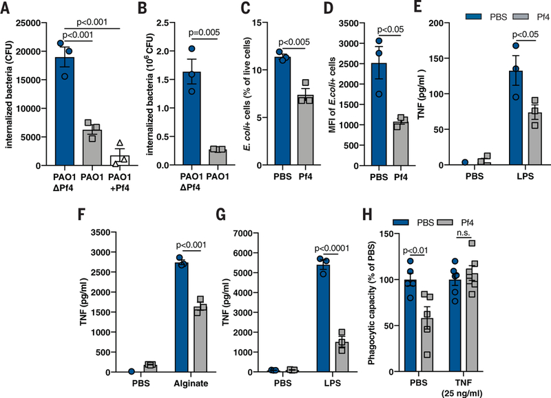
Fig. 2. Pf phage inhibit phagocytosis and TNF production.

(A) Phagocytosis of live PAO1ΔPf4, PAO1, and PAO1 supplemented with exogenous Pf4 (PAO1+Pf4) by mouse BMDCs, as measured by a gentamicin protection assay. (B) Phagocytosis of live PAO1ΔPf4 and PAO1 by human U937 macrophages. (C) Phagocytosis by BMDCs of fixed E. coli particles labeled with a pH-sensitive dye (pHrodo) in the absence or presence of purified Pf4, as measured by flow cytometry. (D) Median fluorescence intensity (MFI) of E. coli pHrodo particle-positive cells from (C). (E and F) TNF production by murine BMDCs stimulated with Pf4 and (E) LPS or (F) alginate for 24 hours. (G) TNF production by human primary monocytes stimulated with Pf4 and LPS for 24 hours. (H) Phagocytosis of E. coli-pHrodo particles by BMDCs stimulated with exogenous TNF and Pf4. All graphs are representative of n ≥ 3 experiments and depict mean with SEM of n ≥ 3 replicates. Analysis: two-tailed Student’s t test.
