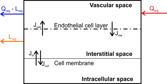
Figure 2.

Structure of an organ in the model. J in, flux into the cell; J ine, flux from the interstitial to the vascular space; J out, flux out of the cell; J vas, flux from the vascular to the interstitial space; L org, regional lymph flow; Q org, regional blood flow.
