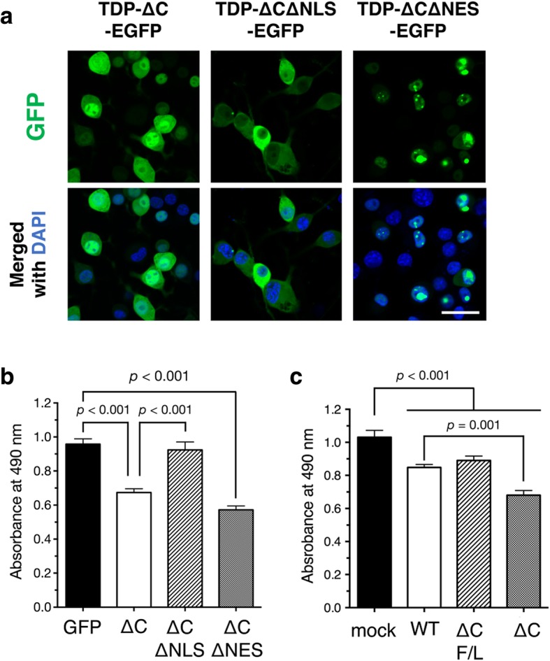
Fig. 4.

Intranuclear TDP-∆C induces cytotoxicity of cultured neuronal cells. a Representative images show subcellular localization of C-terminal EGFP-tagged TDP-∆C (TDP-∆C-EGFP), TDP-∆C with mutations in the NLS (TDP-∆C∆NLS-EGFP), and TDP-∆C with mutations in the NES (TDP-∆C∆NES-EGFP) expressed in mouse neuroblastoma Neuro2a (N2a) cells as well as merged images with DAPI. Scale bar: 20 μm. b and c Nuclear localization (b) and nucleotide binding ability (c) of TDP-∆C are crucial for cytotoxicity. N2a cells were transfected with the indicated expression plasmids and incubated for 48 h in the differentiation media. Cell viability was measured by MTS assay. ∆C F/L represents a TDP-∆C variant with F147 L / F149 L mutations defective in nucleotide binding. Data are expressed as mean ± SEM of three independent experiments, each performed in triplicate
