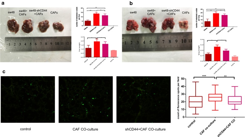
Fig. 5.
CC-CAFs enhanced adhesion and metastasis of CRC cells in vivo. a Liver tissue from BALB/c (nu/nu) mice of each group was obtained at 6 weeks, the number of liver metastatic foci was counted. And weight of liver was quantified. *P < 0.05, **P < 0.01. b Lung tissue from BALB/c (nu/nu) mice of each group was obtained at 8 weeks, the number of lung metastatic foci was counted. And weight of lung was quantified. *P < 0.05, **P < 0.01. c 1 × 10⁶ SW48-GFP cells were injected via tail vein, and 48 h later lungs were taken out for frozen sections. The count of sw48-GFP with or without transfection of shRNA CD44 or pre-co-cultured with CC-CAFs in frozen section per field was analyzed (magnification, ×100). Error bars represent mean ± s.d; **P < 0.01; ***P < 0.001; n.s not significant; by one-way analysis of variance (ANOVA)

