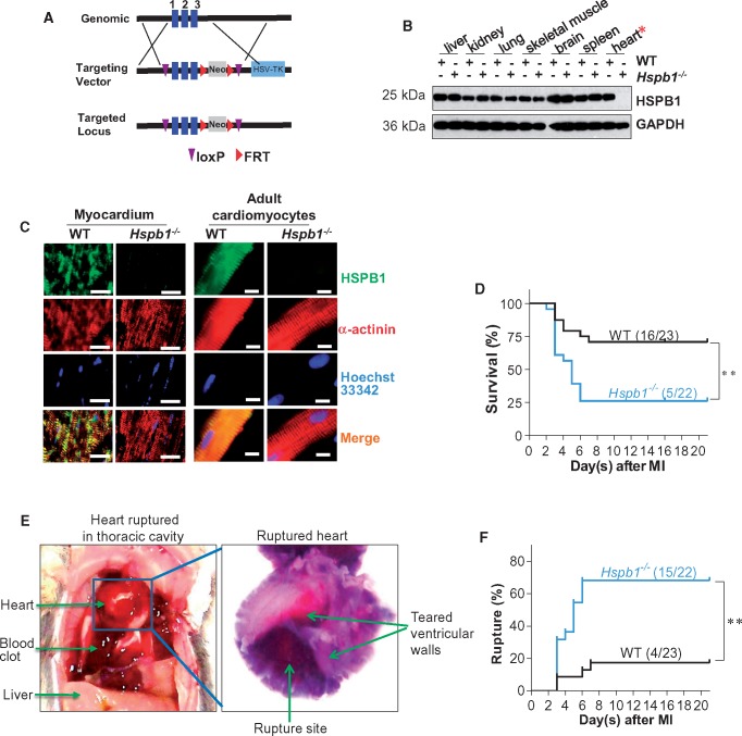
Figure 2.
Hspb1−/− mice showed increased cardiac rupture and mortality after MI. (A) Strategy for creating HSPB1 conditional mice using loxP recombinant system. (B) Deletion of HSPB1 in Hspb1−/− hearts. Different tissues were collected from 8-week-old WT and Hspb1−/− mice. HSPB1 protein expression was analysed using immunoblotting. Note that HSPB1 was absent in Hspb1−/− hearts. n = 6/group. (C) Deletion of HSPB1 in cardiomyocytes of Hspb1−/− mice. Ventricular tissues (left panel) or isolated adult cardiomyocytes (right panel) were collected from 8-week-old WT and Hspb1−/− mice. Immunostaining against HSPB1 (green) was performed. Alpha-actinin was used to stain cardiomyocytes (red). Hoechst 33342 reagent was used to counterstain the nuclei (blue). Note that HSPB1 expression was absent in cardiomyocytes of Hspb1−/− mice. Scale bars, 10 μm. n = 6/group. (D) Kaplan–Meier survival curves. Hspb1−/− and WT mice were subjected to MI insult. Mice survival was recorded within 21 day post-MI. **P < 0.01 analysed by log-rank test. n = 22–23 per group. (E and F) Cardiac rupture. Autopsy was performed in MI-induced death of mice. Cardiac rupture was characterized by presence of intrathoracic blood clots (left panel, E) and perforation on the infarcted wall (right panel, E). Note that a significant more incidence of cardiac rupture was detected in Hspb1−/− mice after MI (F). **P < 0.01 analysed by log-rank test. n = 22–23 per group.

