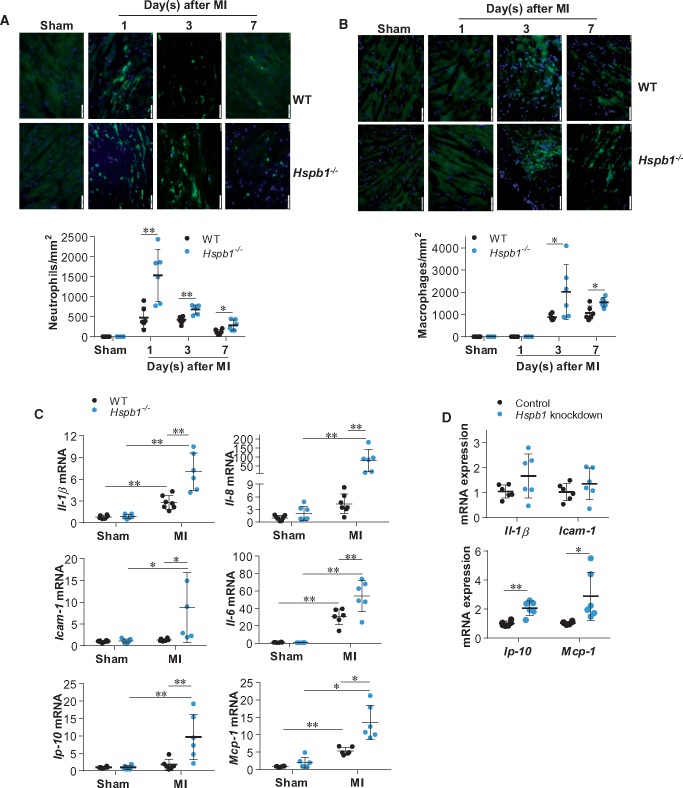
Figure 5.
Deficiency of HSPB1 enhanced the MI-induced inflammatory responses and chemokine induction in cardiomyocytes. (A and B) Leucocyte recruitment. Ventricular tissues at the papillary muscles level were collected after MI for the indicated times. Cryosections were prepared for immunofluorescence staining against neutrophil (A) or F4/80 (B) (green). Hoechst 33342 reagent was used to counterstain the nuclei (blue). Data were expressed as mean ± SD and analysed using unpaired t-test. *P < 0.05, **P < 0.01, n = 6/group. Scale bars, 50 μm. (C) Cytokines and chemokines in myocardium. Ventricular tissues were collected 24 h post-MI for mRNA analysis. Data were expressed as mean ± SD and analysed using two-way ANOVA followed by post hoc test. **P < 0.01 and *P < 0.05, n = 6–8 per group. (D) Cytokines and chemokines in cardiomyocytes. Primary cardiomyocytes were transfected with Hspb1 siRNA to knock down HSPB1 expression. Cardiomyocytes transfected with scramble RNA served as controls. Following hypoxia for 6 h, cardiomyocytes were harvested for mRNA analysis. Data were expressed as mean ± SD and analysed using unpaired t-tests. **P < 0.01 and *P < 0.05, n = 6/group.

