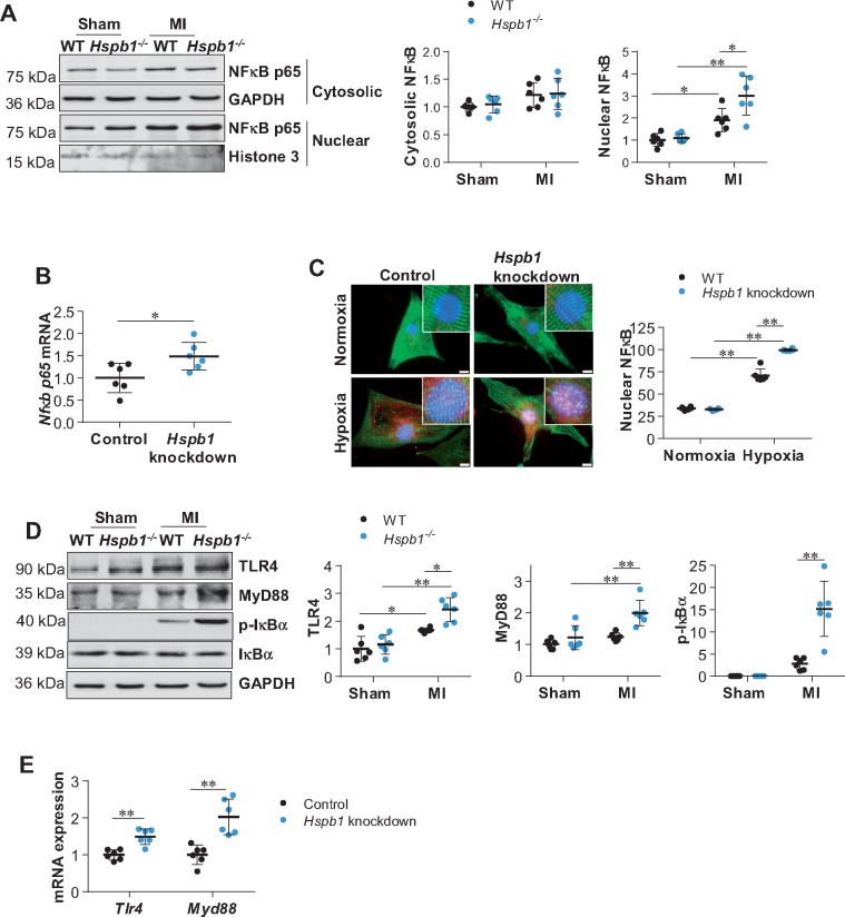
Figure 6.
Deficiency of HSPB1 augmented the MI-induced NFκB activation in cardiomyocytes. (A) NFκB p65 nuclear translocation in myocardium. Ventricular tissues were collected 24 h post-MI. Cytosolic and nuclear protein fractions were prepared for immunoblotting against NFκB p65 subunit. Data were expressed as mean ± SD and analysed using two-way ANOVA followed by post hoc test. **P < 0.01, *P < 0.05, n = 6/group. (B) Nfκb p65 mRNA expression in primary cardiomyocytes. Following hypoxia for 6 h, cardiomyocytes were harvested for analysing Nfκb p65 mRNA levels. Data were expressed as mean ± SD and analysed using unpaired t-tests. *P < 0.05, n = 6/group. (C) NFκB p65 nuclear translocation in primary cardiomyocytes. Following hypoxia for 6 h, cardiomyocytes were harvested for immunostaining against NFκB p65 (red). Alpha-actinin was used to stain cardiomyocytes (green). Hoechst 33342 reagent was used to counterstain the nuclei (blue). Scale bars, 10 μm. Data were expressed as mean ± SD and analysed using two-way ANOVA followed by post hoc test. **P < 0.01, n = 6/group. (D) TLR4/MyD88 signalling in myocardium. Ventricular tissues were collected 24 h post-MI for immunoblotting analysis. Data were expressed as mean ± SD and analysed using two-way ANOVA followed by post hoc test. **P < 0.01, *P < 0.05, n = 6/group. (E) TLR4/MyD88 signalling in cardiomyocytes. Following hypoxia for 6 h, cardiomyocytes were harvested for mRNA analysis. Data were expressed as mean ± SD and analysed using unpaired t-test. **P < 0.01, n = 6/group.

