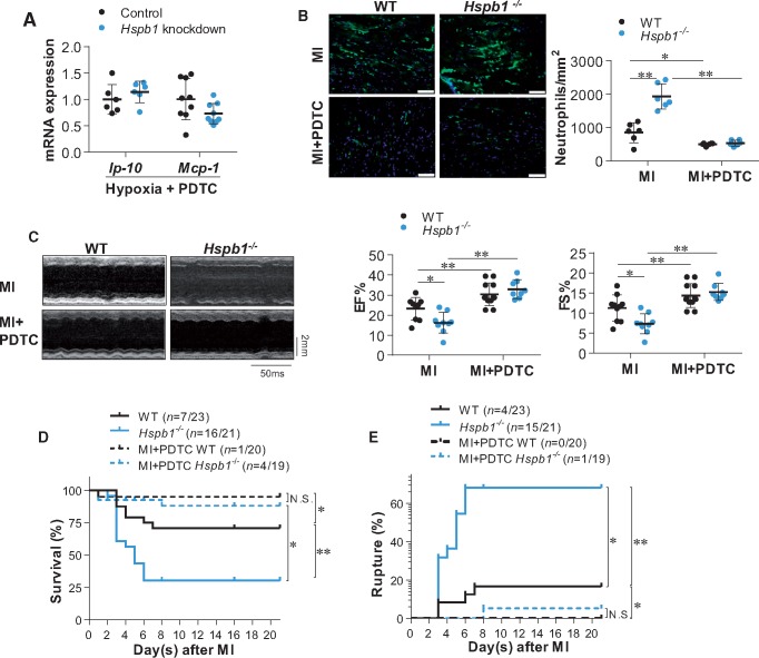
Figure 7.
Inhibition of NFκB reversed the HSPB1 deficiency-induced exaggeration of cardiac damages after MI. (A) Chemokine expression in cardiomyocytes. Cardiomyocytes were exposed to hypoxia for 6 h in the presence of PDTC. Levels of mRNA were examined. Data were expressed as mean ± SD and analysed using unpaired t-test. n = 6–9/group. (B) Neutrophil infiltration in myocardium. Ventricular tissues at the papillary muscles level were collected at 24 h post-MI in the presence or absence of PDTC. Cryosections were prepared for immunofluorescence staining against neutrophil (green). Hoechst 33342 reagent was used to counterstain the nuclei (blue). Data were expressed as mean ± SD and analysed using two-way ANOVA followed by post hoc test. Scale bars, 50 μm; **P < 0.01 or *P < 0.05, n = 6/group. (C) Cardiac function. Cardiac function was evaluated using echocardiography 24 h post-MI in the presence or absence of PDTC. Data were expressed as mean ± SD and analysed using two-way ANOVA followed by post hoc test. Time stamp and scale bar of M-mode images were shown in the figure. **P < 0.01 or *P < 0.05, n = 8–12/group. (D) Kaplan–Meier survival curves. Mice survival was recorded within 21 day post-MI in the presence or absence of PDTC. **P < 0.01 or *P < 0.05 analysed using log-rank test. n = 19–23/group. N.S., no significance. (E) Cardiac rupture. Autopsy was performed to examine cardiac rupture in MI-induced death of mice in the presence or absence of PDTC. **P < 0.01 or *P < 0.05 analysed using log-rank test. n = 19–23/group. N.S., no significance.

