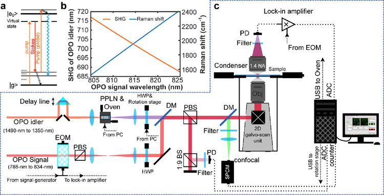
Fig. 1. New stimulated Raman excited fluorescence (SREF) laser microscopy system opening excitation band near 600-nm.
(a) Energy diagram for SREF process. (b) Wavelength of SHG of OPO idler (red) and corresponding resonance Raman shift (blue) as a function of OPO signal wavelength when OPO signal is set as Stoke beam and the SHG is set as pump beam. (c) System setup for SREF excitation band around 600-nm. PPLN, periodically poled Lithium Niobate; QWP, quarter-wave plate; DM, dichroic mirror; PBS, polarization beam splitter; BS, beam splitter; SPCM, single photon counting module; DAC, digital-to-analog converter; ADC, analog-to-digital converter; EOM, electro-optical modulator; APD, avalanched photodiode; PD, photodiode.

