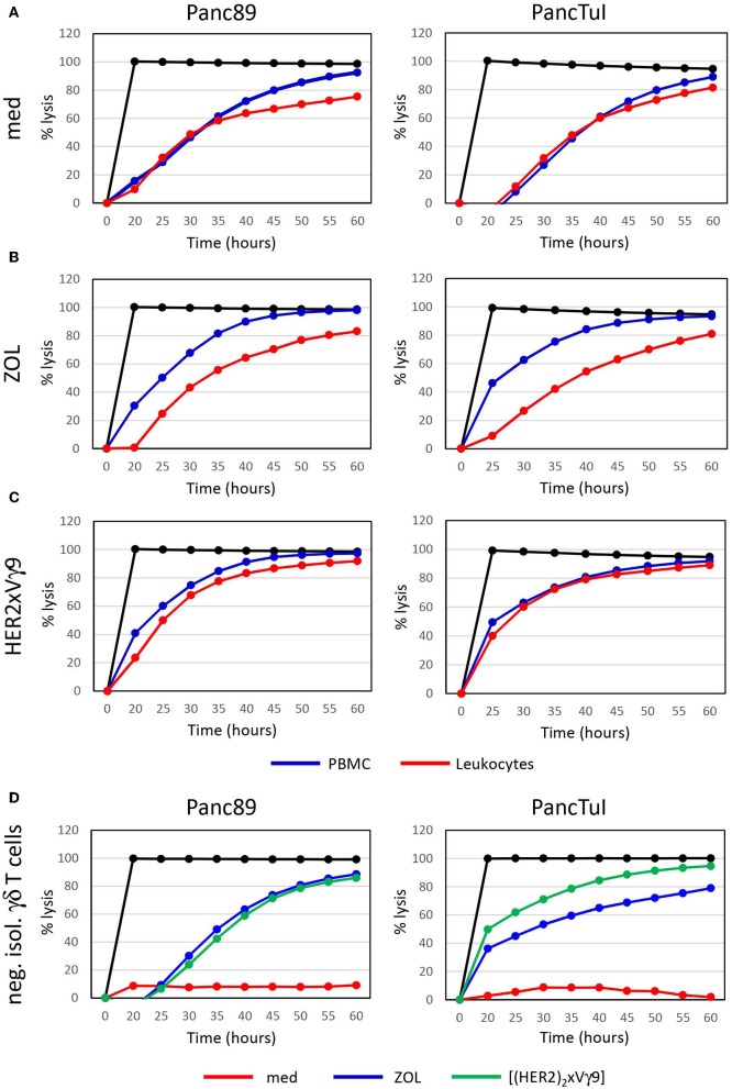
Figure 1.
Zoledronic acid-stimulated leukocytes diminish γδ T cell-cytotoxicity against PDAC cells. (A–C) Cytotoxicity of 250 × 103 PBMC (blue line) or 500 × 103 leukocytes (red line), which each comprised ~7,500 γδ T cells or (D) 125,000 negatively isolated, resting γδ T cells (neg. isol. γδ T cells) in co-cultures with Panc89- (left panel) or PancTuI cells (right panel). Co-cultures were set up in rIL-2 medium only (A), or were additionally stimulated with (B) zoledronic acid (ZOL, 2.5 μM) or (C) with bispecific Ab (bsAb) ([(HER2)2×Vγ9], 1 μg/mL). Cytotoxictiy was analyzed by RTCA. 5 × 10³ PDAC cells were seeded 24 h before addition of effector cells. Percentage of lysis (“% lysis”) was analyzed from RTCA data by calculating the normalized impedance of spontaneous lysis (cell growth of tumor cells in medium alone) in relation to the maximal lysis induced by 1% Triton-X-100 (black line) at indicated time points. Shown is one representative experiment with the same donor out of three independent experiments with different donors.

