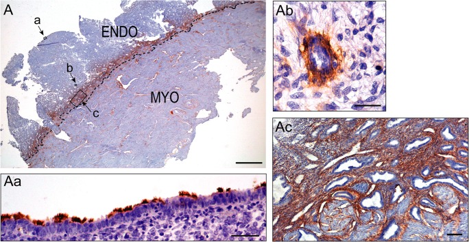
Fig. 1.
Immunolocalization of NTPDase2 in a human secretory endometrium (Endo) and myometrium (Myo) by immunohistochemistry. Image A shows the immunolabeling of NTPDase 2 at the endometrial stroma but only of the basal layer. Image Aa shows the presence of NTPDase2 in the cilia of ciliated epithelial cells of the surface epithelium. Ab is a detail of the NTPDase2 immunolabeling in cells in a perivascular location. Ac shows a detail of the basal layer. NTPDase2 antibody used was H9s from http://ectonucleotidases-ab.com. Scale bars 2 mm (A), 50 μm (Aa), 25 μm (Ab), and 100 μm (Ac)

