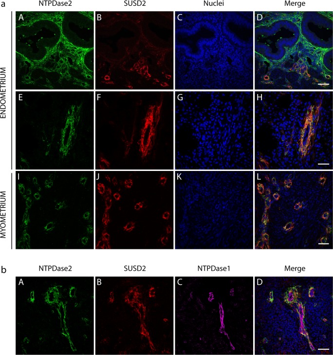
Fig. 4.
a Confocal fluorescence images of a secretory (A–D) and a proliferative (E–H) endometrium and myometrium (I–L) labeled with the antibodies against NTPDase2 (A, E, I) and the eMSC marker SUSD2 (B, F, J). Nuclei were labeled with DAPI (C, G, K). The images A–D show part of the basal layer of a secretory endometrium. NTPDase2 label is present in cilia of ciliated cells of glands, in stromal cells, especially periglandularly, and in perivascular cells. Images E–H show a detail of an endometrial vessel localized in the functional layer of a proliferative endometrium and images I–L show a detail of the vessels in myometrium. NTPDase2 is only present in perivascular cells (E, I). The eMSC marker SUSD2 is present in perivascular cells (B, F, J). Merge images (D, H, L) show the colocalization of NTPDase2 and SUSD2 in the perivascular cells. Scale bars 50 μm (D, H) and 35 μm (L). b Immunolocalization of NTPDase2 (A), SUSD2 (B), and NTPDase1 (C) in the vessel of an atrophic endometrium. Nuclei were labeled with DAPI. NTPDase2 (A) and SUSD2 (B) were present in perivascular cells while NTPDase1 was detected in endothelial cells (C). Merged image shows the colocalization of NTPDase2 and SUSD2 in perivascular cells (D). Scale bar 50 μm (D) NTPDase2 antibody used in all the experiments shown in this figure was H9s from http://ectonucleotidases-ab.com

