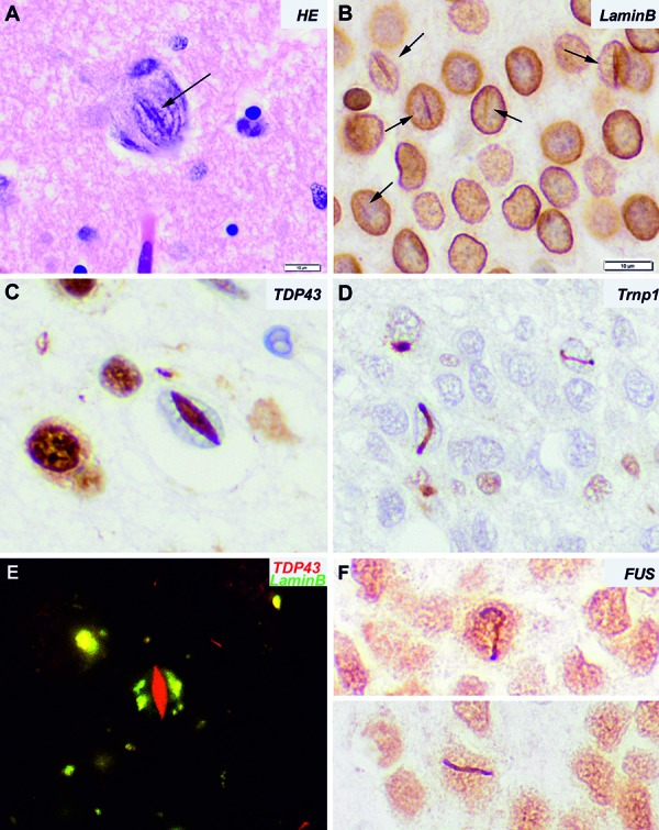
Figure 1. A: Hematoxylin-Eosin staining of a large neuron with a central basophilic cleft crossing the nucleus. B: Immunohistochemistry for lamin B1 nicely depicts the nuclear envelop and enhances the nuclear clefts. C: Cat-eye lentiform intranuclear inclusion in a patient with FTLD-TDP. D: Immunohistochemistry for Transportin 1 shows small lanceolated or vermiform intranuclear inclusions in granular neurons of the dentate gyrus of the hippocampus in a patient with aFTLD-U/ FTLD-FET. These inclusions are also immunoreactive for FUS (F). E: Double immunofluorescence for TDP43 and lamin B1 reveals that the cat-eye inclusion is not surrounded by the nuclear membrane. Instead, lamin B1 immunoreactivity shows an irregular distribution throughout the nucleus.

