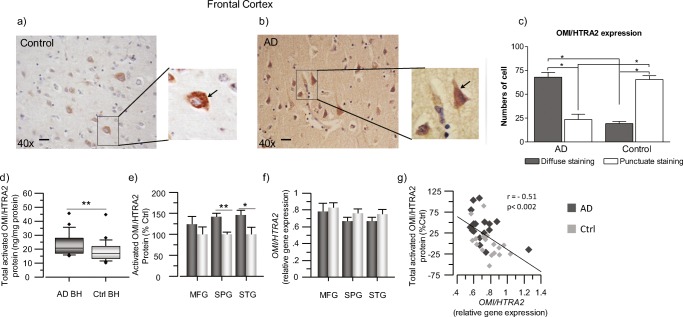
Fig. 1.
OMI/HTRA2 protein level is higher in AD than control brains. a, b Immunohistochemical detection of OMI/HTRA2 in human postmortem brain sections of frontal cortex. Representative image of sections from non-demented controls (a) and AD (b) subjects. Staining: OMI/HTRA2 (brown): anti-OMI/HTRA2 rabbit primary antibody, biotinylated horse anti-rabbit antibody, and ABC-Elite HRP. Counterstaining: hematoxylin (blue). Scale bar = 20 μm. c Quantitative analyses showing the number of cells that present either a diffuse or punctuate staining pattern of OMI/HTRA2 in postmortem brain tissues. d, e Quantification of activated OMI/HTRA2 protein levels in postmortem brain homogenates (BH) of AD subjects (n = 6) and non-demented controls (n = 6), by sandwich ELISA. d Box plot graph showing the overall level of activated OMI/HTRA2 in AD compared to controls, measured in all the extracts from the three brain regions and expressed as ng/mg of total protein. e Graph showing the level of activated OMI/HTRA2 in each brain region separately, expressed as percentage of the control levels. f, gOMI/HTRA2 gene expression was quantified in the postmortem brain samples from the MFG, SPG, and STG brain regions. f Graph showing the normalized transcript levels of OMI/HTRA2 in each of the analyzed brain regions. g Negative correlation of OMI/HTRA2 transcript levels and activated OMI/HTRA2 protein levels, expressed as % of the mean of the controls (Ctrl). Activated OMI/HTRA2 was quantified in medial frontal gyrus (MFG), superior temporalis gyrus (STG), and superior parietal gyrus (SPG) brain regions. The total activated OMI/HTRA2 refers to the sum of its protein levels in all extracts. AD, dark gray squares; non-demented controls, light gray squares. Mean values ± S.E.M. are shown (two asterisks, p < 0.01; one asterisk, p < 0.05)

