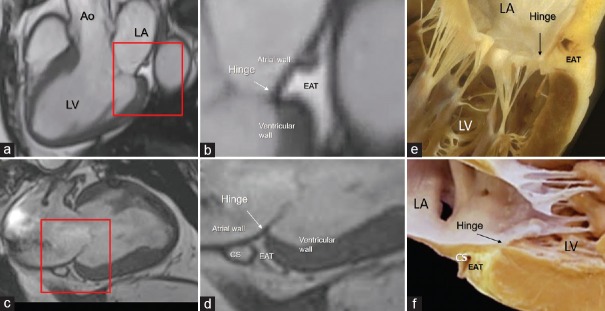
Figure 6.
CMR SSFP still frame in long axis (a and b) and in 2 chamber (c and d) views. The areas in the red box in the panels a and c are magnified in panels b and d. The images clearly show the EAT occupying the atrioventricular groove up to the base of the posterior leaflet insertion. e and f are anatomic specimens showing the hinge (arrow) of the mitral leaflet interposing between atrial and ventricular walls and the yellow colored EAT. Ao = Aorta, LA = Left atrium, LV = Left ventricle, CS = Coronary sinus, CMR SSFP = Cardiac magnetic resonance steady-state free precession, EAT = Epicardial adipose tissue

