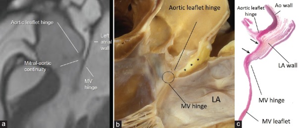
Figure 8.
(a) CMR SSFP still frame in long axis view showing in patient with abundant EAT as the adipose tissue separates the aortic wall from the atrial wall in the transverse pericardial sinus. Panel (b) shows anatomic specimen displayed in similar view showing EAT (asterisk) between the outside of the aortic sinus and the anterior wall of the LA. Aortic-mitral fibrous continuity is circled. Panel (c) is a histologic section in similar orientation to show fibrous tissue (stained red between the two arrows). The LA wall (stained brownish) is on the outside but there is no ventricular muscle in between aortic and mitral leaflets (see text). LA = Left atrium, CMR SSFP = Cardiac magnetic resonance steady-state free precession, EAT = Epicardial adipose tissue

