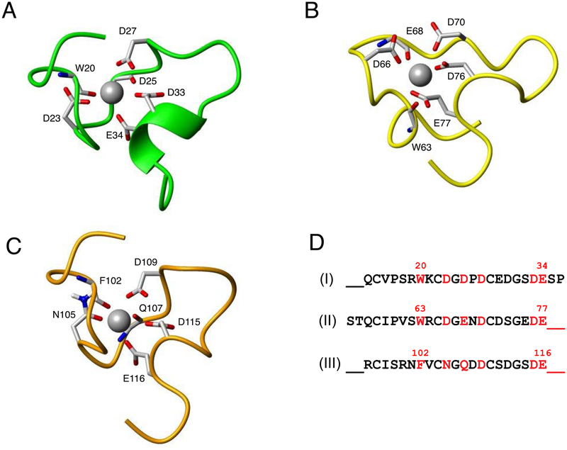
Figure 6.
Calcium-binding pockets of the VLDLR(2–4) fragment. Ligands for Ca2+ binding in CR2, CR3, and CR4 domains are shown in (A), (B), and (C), respectively. Ca2+ ions are shown as grey spheres, while side chains of the residues that co-ordinate with these ions are shown in sticks. The structures of the three calcium-binding loops are shown by superimposing them on the last three residues (S32-E34, E75-E77, S114-E116) so that they have the same relative orientation. (D) Sequence alignment of the three Ca2+-binding regions of CR2, CR3, and CR4 domains (I, II, and III, respectively) with the ligand residues colored in red and the 2-residue inserts in CR2 and CR3 shown at the end and the beginning of their sequences, respectively.

