Figure 1.

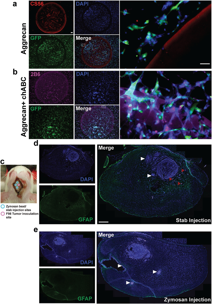
Astroglial scar constituents repel brain tumor cells. a) An in vitro model of glial scar was implemented using a spot assay with the CSPG, aggrecan. U87mg GBM cells expressing green fluorescent protein (GFP) were used. DAPI indicates the nuclear stain 4’,6-diamidino-2-phenylindole, and CS56 is an antibody that stains intact CSPGs. A spot of CS56+ aggrecan (1 mg mL−1, 2 μL spot) repels the tumor cells. Dotted line indicates zoomed in region on right, confirming that tumor cells cannot cross the boundary posed by aggrecan. b) Enzymatic digestion of CSPG GAGs abolishes CSPG-mediated tumor cell invasion inhibition. 2B6 is an antibody that stains the GAG stubs following enzymatic digestion of aggrecan by chondroitinase ABC (chABC). Tumor cells were able to cross the spot boundary posed by aggrecan upon chABC digestion indicating that CSPGs mediate their repulsive effects via CS-GAGs. c) Schematic of coinjection of zymosan beads (or control stab wounds in separate animals) and F98 tumors in Fischer rats. d) Glial fibrillary acidic protein (GFAP) is an immunofluorescent stain for reactive astrocytes. Stab wounds, as indicated by white arrow heads partly repelled tumor growth but were unable to prevent tumor microsatellite migration as indicated by red arrow heads. e) Zymosan beads caused fulminant gliosis and cavitation (white arrow heads) and caused tumors to remain as compact masses. Scale bar in (a) is 50 μm, zoomed in region is 200 μm and in (d) is 200 μm.
