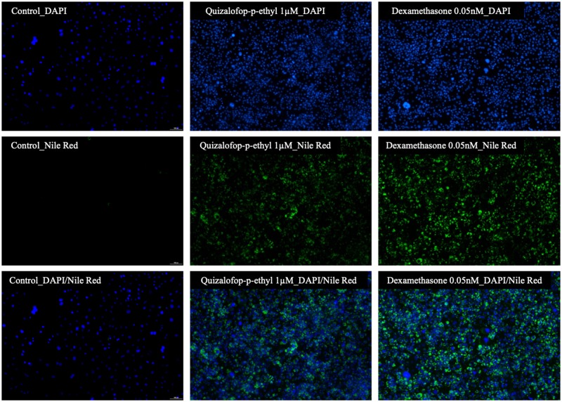
Figure 3.
The adipogenic effect of QpE confirmed using fluorescence imaging. The 3T3-L1 adipocyte cells were cultured with standard treatment-free medium (control), or in the presence of 1 µM QpE or 0.05 nM dexamethasone (positive control). After 8 days of differentiation with the indicated treatments, lipid accumulation was visualized by fluorescent Nile Red staining and the cells visualized by fluorescence microscopy. Nile Red staining for lipid droplets and DAPI staining for cell nuclei were imaged at 530 and 405 nm, respectively, using fluorescence imaging on a Nikon Eclipse Ts2 microscope (40× objective).

