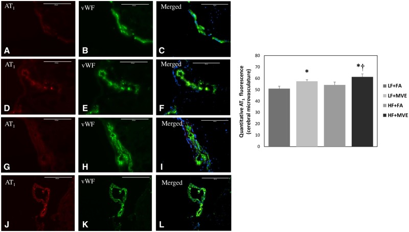
Figure 2.
Double immunofluorescence of angiotensin II type 1 receptor (AT1, A, D, G, J) and vWF (B, E, H, K) in the cerebral microvasculature of C57Bl/6 mice exposed to filtered air (FA) on a low-fat diet (A–C); mixed vehicle emissions (MVE; 100 μg/m3 PM of mixed gasoline and diesel engine emissions) for 6 h/d, for 30 days on a LF diet (D–F); FA+HF diet (G–I) or MVE + HF diet (J–L). n = 3–4 per exposure group; 4 sections each, 3–5 vessels per section. Right panels (C, F, I, L) are merged figures of panels for AT1 and vWF. Blue fluorescence is Hoechst stained nuclei. Scale bar = 100 μm. Results represent mean ± SEM. *p < .05 compared with LF+FA. †p < .05 compared with HF+FA.

