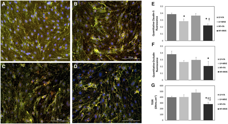
Figure 3.
Representative images of tight junction (TJ) proteins, claudin-5 (green) and occludin (red), fluorescence on endothelial cells (ECs) treated with plasma from mice exposed to (A) filtered air (FA) on a low-fat (LF) diet, (B) exposed to mixed vehicle exhaust (MVE: 100 µg/m3 PM) for 6 h/d, for 30 days on a LF diet, (C) FA + high-fat (HF) diet, or (D) MVE+ HF diet. The cells were fixed and stained for TJ proteins at 24 h post-plasma treatment. Fluorescence was quantified and represented as mean ± SEM (E, F) from n = 3. Blue fluorescence = Hoechst stained nuclei. Scale bar = 100 µm. Trans endothelial electrical resistance measurements in BBB coculture treated with a 1:20 dilution of mouse plasma on the EC (apical) layer from C57Bl/6 mice (G). Transendothelial electrical resistance values were measured 24 h post-plasma treatment. *p < .05 compared with LF+FA. †p < .05 compared with HF+FA. ‡p < .05 compared with LF+MVE.

