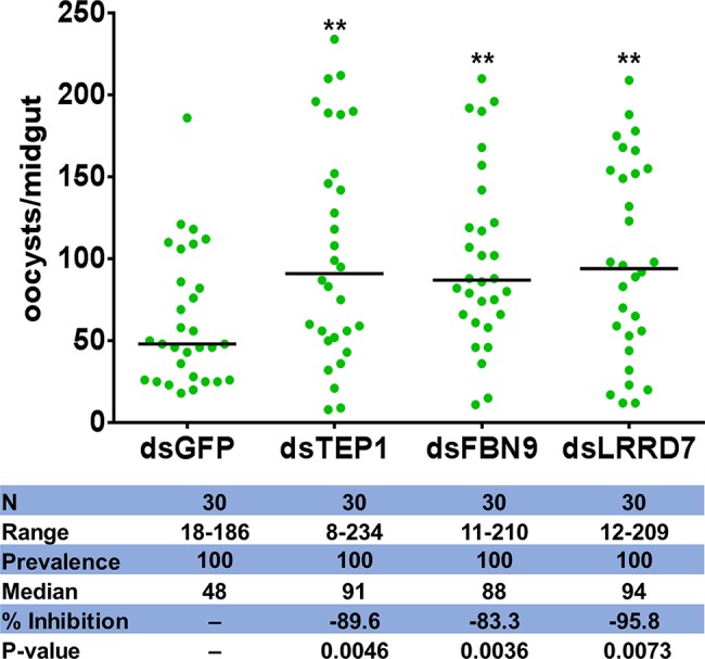
Figure 6.

Oocyst loads following silencing of TEP1, FBN9 and LRRD7 genes. An. stephensi female mosquitoes were injected with dsRNA solution for TEP1, FBN9, LRRD7 or GFP as a control. Injected mosquitoes were colonized with Serratia Y1 and then fed on a P. berghei infected mouse 3 days after dsRNA injection. Mosquito midguts were dissected 10 days post-blood feeding to determine the number of oocysts per mosquito. Each dot represents the number of oocysts from individual midguts, and the horizontal lines indicate the median number of oocysts. The experiments were repeated three times with similar results (Supplementary Figure S8). The Mann-Whitney test was used to determine significance in oocysts numbers, **p < 0.01.
