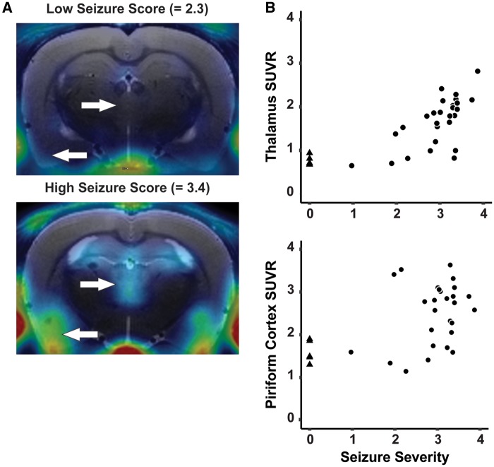
Figure 6.
The extent of neuroinflammation detected by [18F]PBR111 PET is highly correlated with seizure severity. A, Parametric images of SUVR from DFP-intoxicated animals with low versus high SSAVR over the first 4 h postexposure. White arrows indicate differential [18F]PBR111 uptake in the thalamus (arrow facing right) and the piriform cortex (arrow facing left). B, Scatter plot depicting the high positive correlation between SSAVR and SUVR in the thalamus (rs = 0.84; p < .001) and piriform cortex (rs = 0.48; p < .01). DFP-intoxicated animals are indicated with black circles, while VEH control animals are indicated as black triangles.

