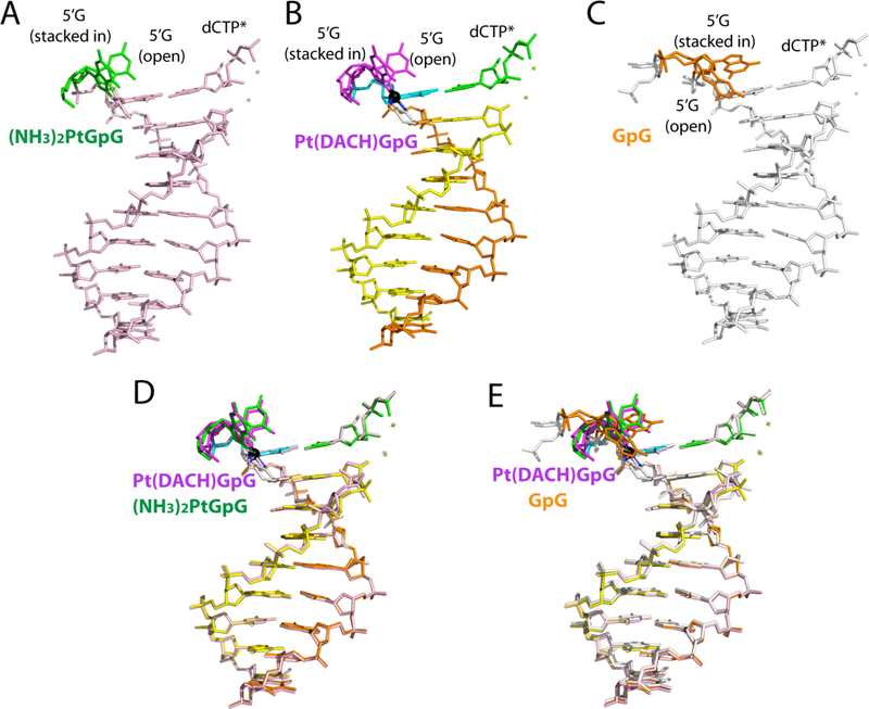
Figure 7. Conformations of DNA in Polη structures.

Comparison of DNA conformations in (A) the Pt(NH3)2–3′G・dCTP* (PDB ID: 4DL4 [39]); (B) the Pt(DACH)−3′G・dCTP*; and (C) the normal-3′G・dCTP* (PDB ID: 4DL2 [39]) structures. Superposition of (D) the Pt(DACH)−3′G・dCTP* and Pt(NH3)2–3′G・dCTP* structures and (E) the Pt(DACH)−3′G・dCTP* and normal-3′G・dCTP* structures.
