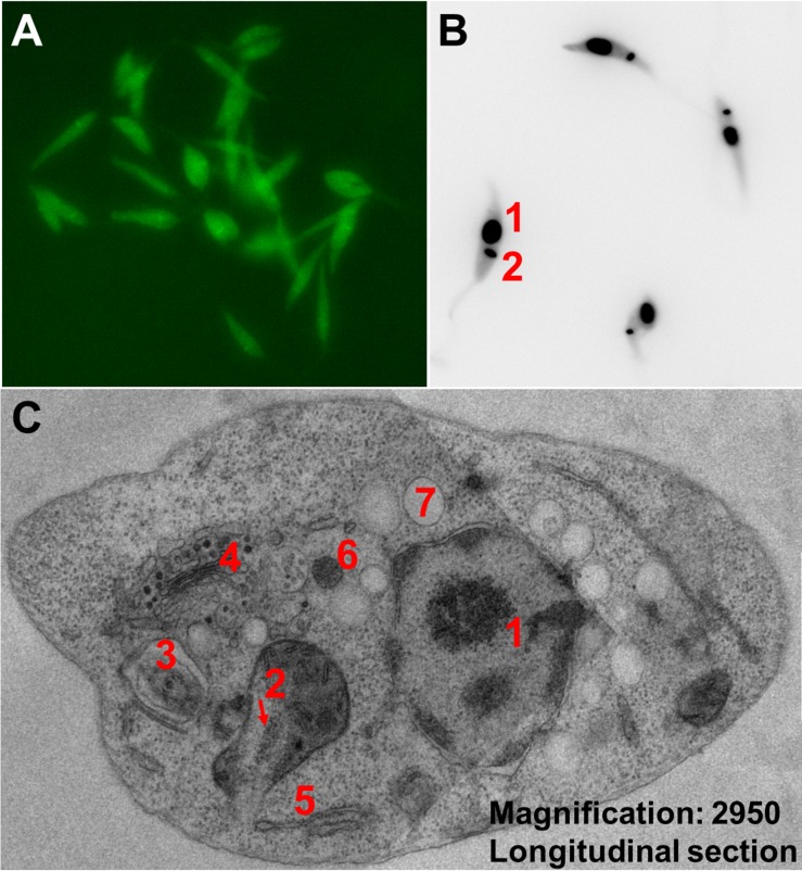
Fig 4. Microscopy images of L. tarentolae.
(A) Fluorescent L. tarentolae promastigotes expressing eGFP. (B) L. tarentolae promastigotes stained with DAPI, highlighting the nucleus (1) and the kDNA (2). (C) TEM (two fused images) of an L. tarentolae promastigote, longitudinal cell section, with a magnification of ×2,950. Cell nucleus (1), kinetoplast inside single mitochondrion (2), flagellum within flagellar pocket (3), Golgi apparatus (4), rough endoplasmic reticulum (5), glycosome (6), and acidocalcisome (7). eGFP, enhanced green fluorescent protein; kDNA, kinetoplastid DNA; TEM, transmission electron microscopy.

