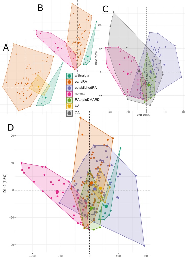
Fig 1. The first two principal components of the PCA based on the RPKMs of the coding genes.
The areas are the convex hulls of the conditions. The largest point of one color depicts the center of a hull. A, B, and D are the same PCA analysis with the same coordinates, where in D all conditions except OA are visible, in A and B only three of them for a better overview. C is a PCA with OA, where four conditions are shown to depict the variability of OA. Number of samples: 10 arthralgia, 57 earlyRA, 95 establishedRA, 27 normal, 22 OA, 19 RAtripleDMARD and 6 UA.

