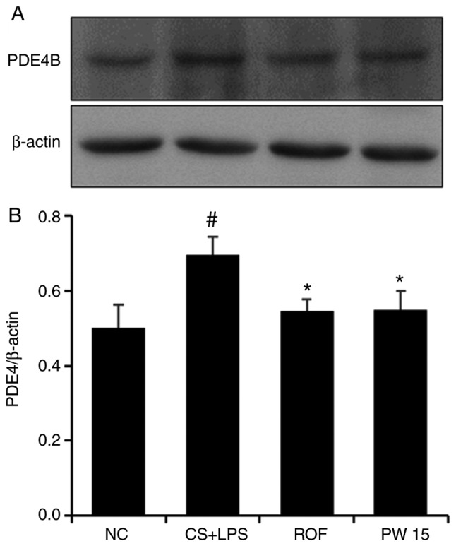
Figure 9.

Effect of PW on PDE4 expression in the lungs of mice with CS- and LPS-induced pulmonary inflammation. (A) Western blot analysis was used to determine the levels of PDE4 in the lung tissue sample. (B) Quantitative analysis of PDE4 expression was performed by densitometric analysis. Data are presented as the mean ± standard deviation (n=6). #P<0.05 vs. NC group. *P<0.05 vs. CS+LPS group. PDE4, phosphodiesterase 4; CS, cigarette smoke; LPS, lipopolysaccharide; PW, Pistacia weinmannifolia root extract; NC, normal control mice; CS+LPS, mice exposed to cigarette smoke and LPS; ROF, mice administered ROF (5 mg/kg) + CS+LPS; PW 15, mice administered PW (15 mg/kg) + CS+LPS.
