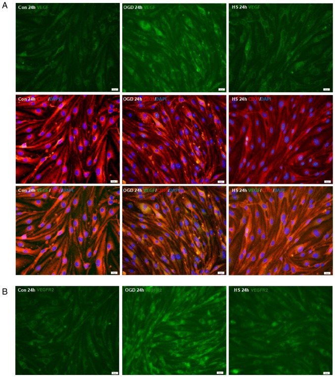
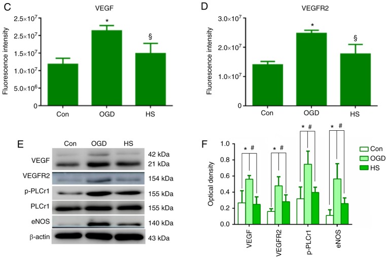
Figure 6.
VEGF, VEGFR2, p-PLCγ1, PLCγ1 and eNOS protein expression in bEnd.3 endothelial cells. (A) Immunofluorescence staining of VEGF (green) in bEnd.3 endothelial cells. Co-localization with the endothelial cell marker CD31 (red) was observed. (B) Immunofluorescence staining of VEGFR2 in bEnd.3 endothelial cells. Scale bar, 20 µm. (C) Fluorescence intensity changes of VEGF protein and (D) VEGFR2 protein in each experimental group. §P<0.05 vs. OGD; and *P<0.05 vs. control. (E) Representative images from western blot analysis for the proteins VEGF (21 and 42 kDa), VEGFR2 (154 kDa), p-PLCγ1 (155 kDa), PLCγ1 (155 kDa), eNOS (140 kDa) and β-actin (43 kDa). (F) Quantification of western blotting results in each group. #P<0.05 vs. OGD; and *P<0.05 vs. control. VEGF, vascular endothelial growth factor; VEGFR2, vascular endothelial growth factor receptor 2; p-, phosphorylated; PLCγ1, phospholipase C γ1; eNOS, endothelial nitric oxide synthase; OGD, oxygen-glucose deprivation; HS, hypertonic saline; Con, control.


