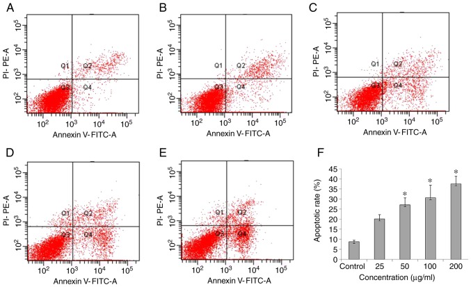
Figure 3.
Effects of SNs on the apoptosis of HL-7702 cells. (A) Cells exposed to basal medium without SNs for 24 h were observed. Cells exposed to (B) 25, (C) 50, (D) 100 and (E) 200 µg/ml of SNs were analyzed via flow cytometry. (F) Apoptotic rates of HL‑7702 cells treated with different concentrations of SNs for 24 h as determined using a flow cytometer. *P<0.05 vs. control. PI, propidium iodide; SN, silica nanoparticle.

